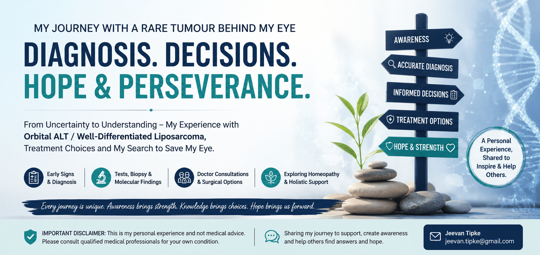
My experience from the first change I noticed in November 2022, through multiple biopsies and scans, identification of tumour behind eye, the recommendation for complete eye removal, and my continuing search for an eye-saving solution
जीवन टिपके
Mumbai, India | Personal account through January 2026
Contact: [email protected]
MEDICAL DISCLAIMER: This is my personal experience, not medical advice. My reports and my doctors’ opinions should be interpreted by qualified specialists using the original records, pathology, molecular tests and imaging. I include my homeopathic treatment because it is part of my story, but I do not claim that homeopathy cured, shrank or stopped my tumour. My symptom improvement and MRI stability do not prove causation. Anyone with a similar condition should seek appropriate ocular-oncology, orbital-surgery, pathology and sarcoma-specialist care and should not delay recommended treatment based on my experience.
1. Why I Am Sharing My Story
I am Jeevan Tipke from Mumbai. I never expected a small change in the appearance of my left eye to become a long medical journey involving steroid treatment, repeated biopsies, MRI scans, PET/CT, molecular testing and discussions about complete orbital exenteration.
I am sharing this story because I found very little information about a rare fatty tumour in the orbit involving an extraocular muscle and lying close to the optic nerve. My goal is to help patients with similar symptoms find information and to make my experience available to doctors and researchers.
2. November 2022 – The First Change I Noticed
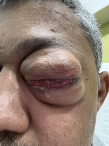
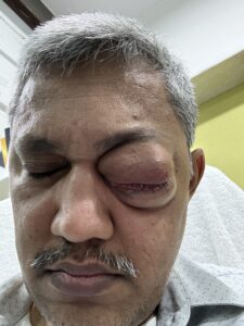
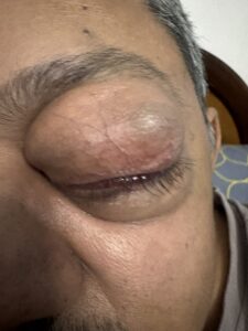
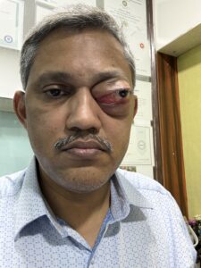
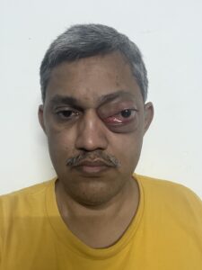
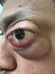
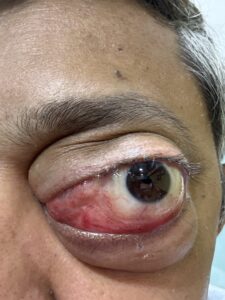
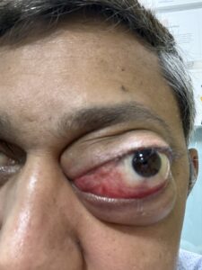
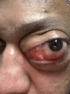
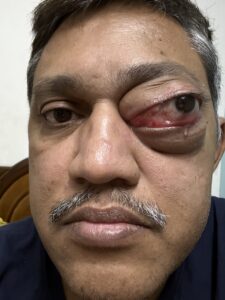
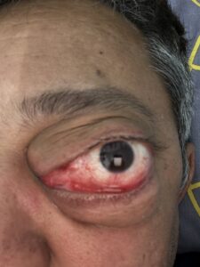
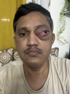
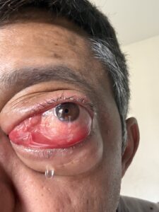
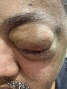
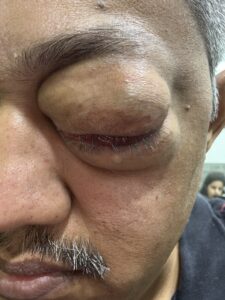
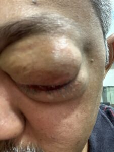
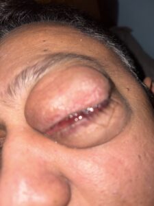
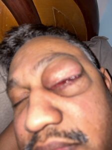
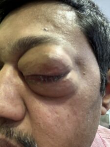
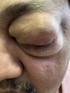
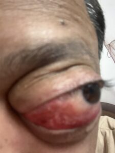
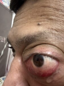
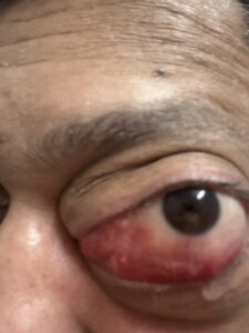
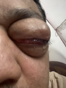
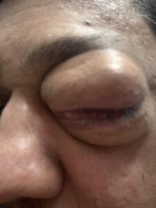
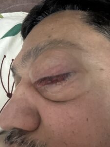
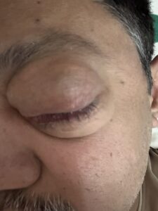
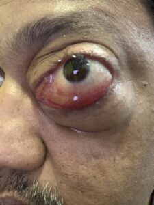
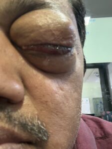
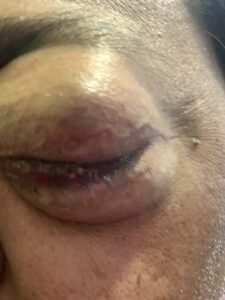
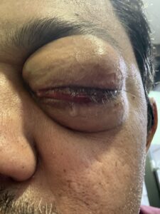
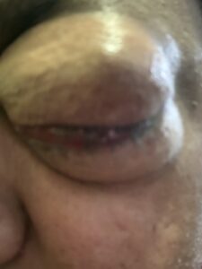
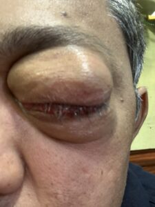
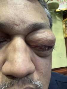
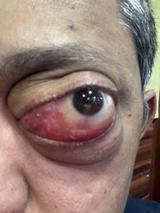
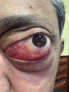
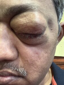
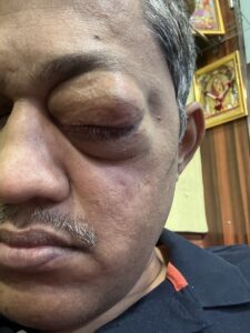
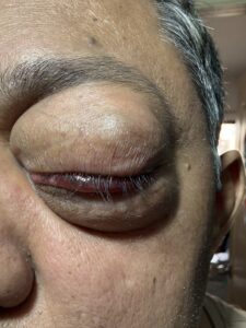
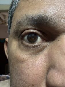
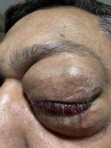
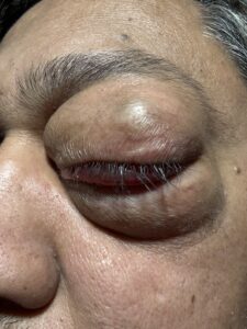
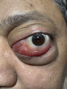
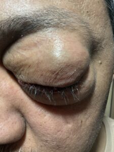
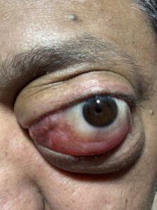
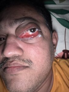
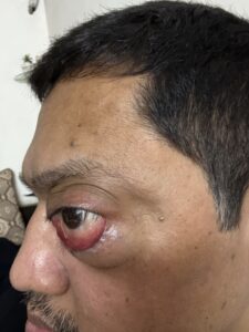
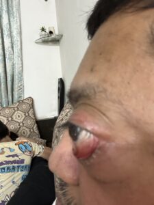
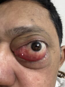
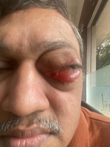
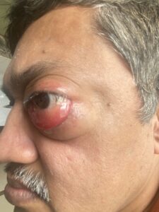
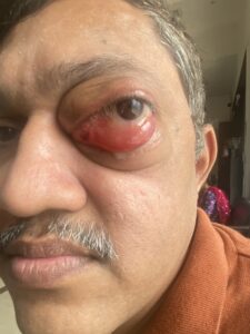
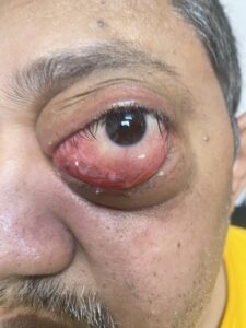
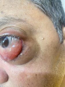
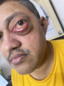
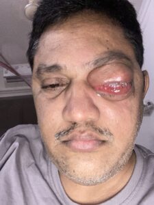
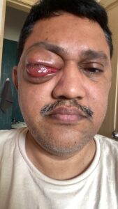
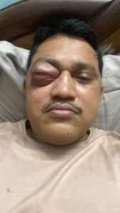
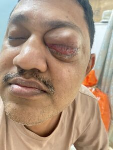
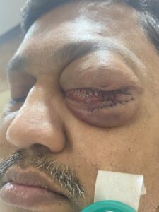
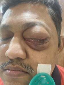
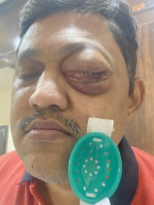
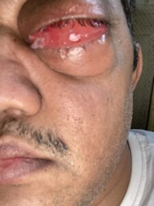
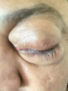
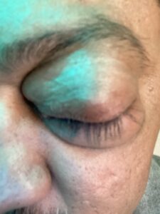
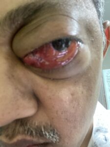
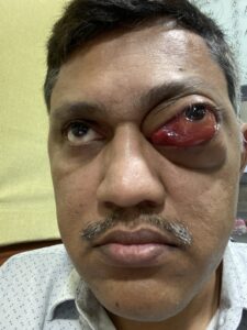
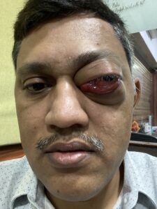
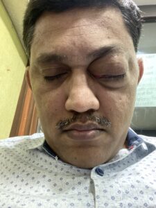
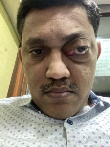
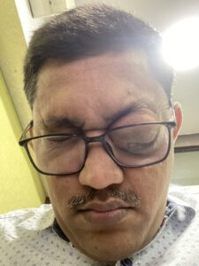
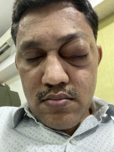
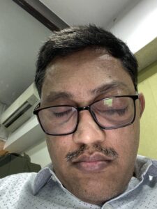
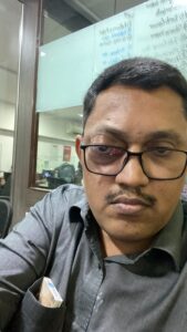
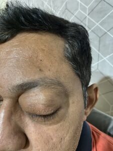
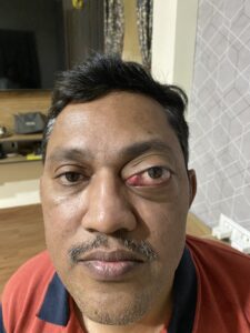
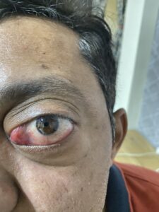
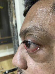
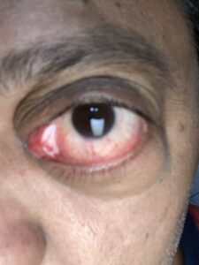
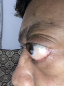
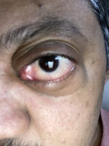
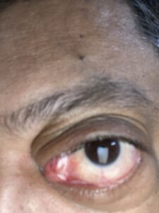
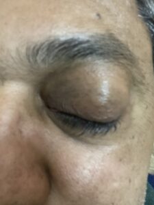
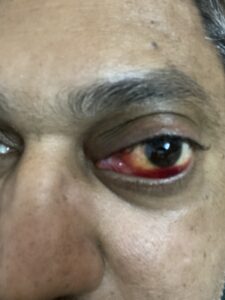
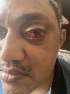
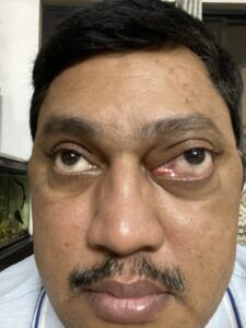
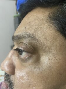
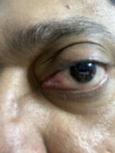
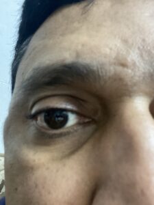
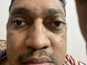
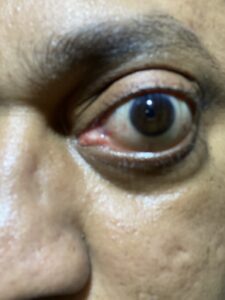
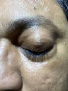
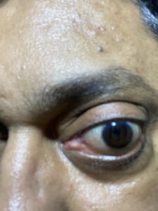
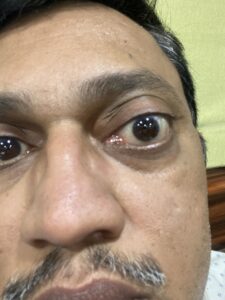
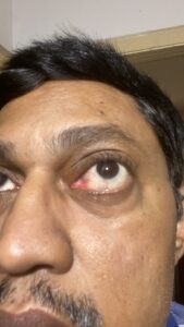
In November 2022, I noticed that my left eye looked slightly larger and more prominent than before. The change was initially small, but it was unusual for me, so I sought an eye consultation.
I visited Laxmi Eye Hospital in Panvel, Navi Mumbai. At that time I had no idea that this would be the beginning of a multi-year investigation into a rare orbital mass.
3. Four Weekly Steroid Injections
Initially, the doctors treated me as though inflammation could be responsible. I received four sets of steroid injections, approximately one injection once a week for four weeks. I was then asked to wait and observe the eye for several weeks.
By around January 2023, the overall eye size had not returned to normal and the eye continued to look slightly enlarged. This made it increasingly important to understand what was actually causing the problem.
4. 2023 – Medicines and Observation
Throughout 2023 I continued the tablets/medicines prescribed by my doctor for the recommended periods, while we continued to observe the eye. The underlying cause was still not clear.
At that stage, the possibilities included an inflammatory or autoimmune orbital condition and other causes of an enlarging orbital lesion.
5. January–March 2024 – My First Biopsy
In January 2024, my doctor recommended a biopsy. I underwent the procedure at a renowned hospital around March 2024. Unfortunately, the biopsy did not identify the root cause.
A second biopsy was subsequently performed, but that too did not provide a definitive answer. This was one of the most difficult parts of my journey: the eye was visibly abnormal, yet the tissue samples had not explained why.
6. Referral to Centre For Sight, Hyderabad
Because the diagnosis remained unresolved, I was advised to consult Centre For Sight in Hyderabad. Dr. Santosh G. Honavar and the team reviewed my case and another biopsy was performed.
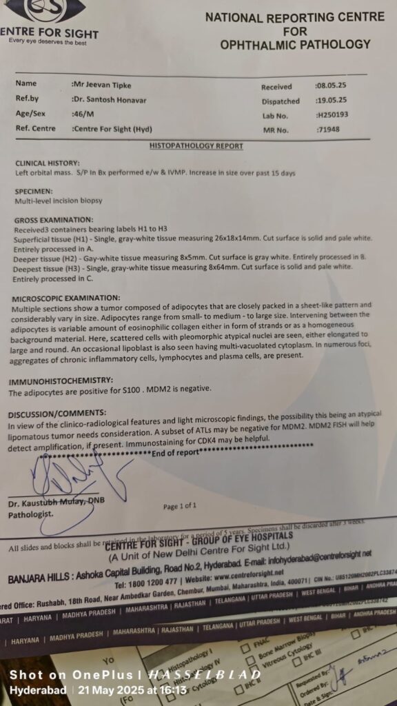
Earlier pathology had suggested a reactive chronic inflammatory lesion, with lymphoid and plasma cells around blood vessels. There was no granuloma or vasculitis and special stains for bacteria and fungi were negative. Idiopathic orbital inflammation and IgG4-related disease were among the possibilities at that stage.
Later pathology and imaging shifted the investigation toward a fatty/lipomatous tumour.
7. What My MRI Showed
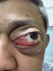
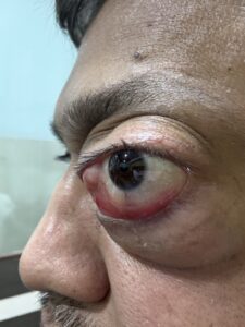
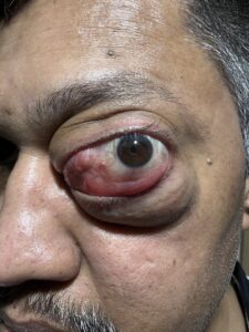
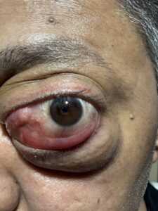
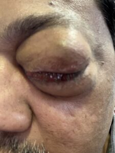
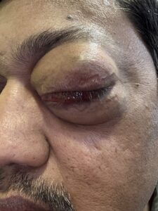
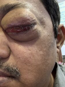
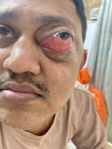
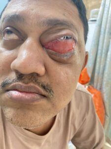
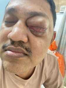
My 9 April 2025 MRI showed gross left-sided proptosis caused by an ill-defined mass in the intraconal and extraconal parts of the orbit. The lesion extended back to the orbital apex.
The left medial rectus muscle was infiltrated and near-totally replaced by the mass. The left optic nerve was displaced, compressed and stretched. The lesion was predominantly fat signal with interspersed soft-tissue components. The radiologist considered liposarcoma and pleomorphic lipoma among the possibilities.
8. PET/CT – Checking for Disease Elsewhere
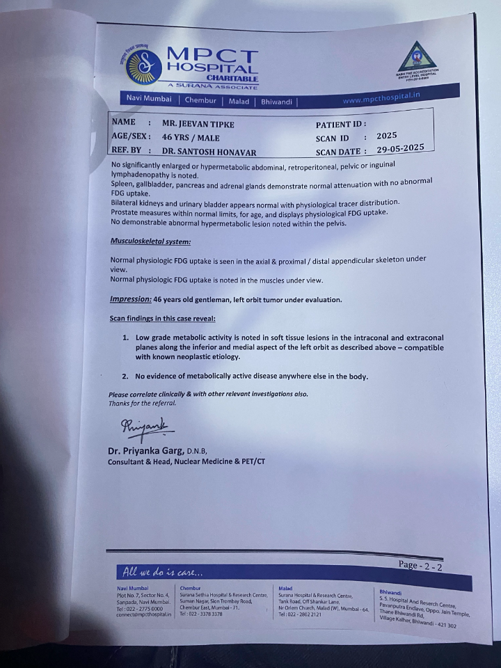
My whole-body PET/CT showed low-grade FDG uptake in the orbital lesion. Importantly, the report did not identify other metabolically active disease elsewhere in the body.
That was reassuring regarding distant disease, but the local orbital tumour remained a serious issue because of its size and relationship to the extraocular muscles and optic nerve.
9. Biopsy, Molecular Testing and the ALT/WDLPS Question
The later investigation led to concern for atypical lipomatous tumour (ALT) / well-differentiated liposarcoma (WDLPS) of the orbit (tumour behind eye). Molecular testing detected CDK4 and MDM2 copy-number gains.
The molecular report also recommended confirmation by an alternate method because of assay limitations. Therefore, I believe the final diagnosis should always be interpreted by an expert pathologist together with the original biopsy material, imaging and any confirmatory MDM2/CDK4 testing.
This complicated diagnostic journey is one reason I am documenting the case in detail.
10. Why Complete Eye Removal Was Recommended to get rid tumour behind eye.
Because the tumour was extensive, involved the medial rectus muscle and was closely associated with the optic nerve and other orbital structures, my doctors recommended complete orbital exenteration/marginal surgery as the most definitive surgical approach.
For me, this was an enormous decision. Losing the eye is irreversible, so I wanted to understand whether an eye-sparing operation, debulking, radiotherapy or another approach might be possible.
11. Tata Memorial Hospital and My Search for Another Opinion
I later visited Tata Memorial Hospital in Mumbai and discussed my case with Dr. Deepa Nair from the Head and Neck department. Their radiology team also reviewed the case.
Because this is rare and the proposed operation is irreversible, I wanted multidisciplinary opinions before making a final decision.
12. Why I Decided to Wait
At that stage I decided to pause before proceeding with complete eye removal. I was not ignoring the seriousness of the disease. I wanted to understand the natural history, recurrence risk, eye-sparing possibilities and whether additional research or treatments could become relevant.
I also wanted objective follow-up rather than simply waiting without medical monitoring.
13. Starting Homeopathy – 30 June 2025
On 30 June 2025, I started homeopathic treatment with Dr. Borkar in Dadar, Mumbai. I communicated this decision to my allopathic doctor.
I am including this in my story because it became part of my treatment journey. I am not presenting homeopathy as a proven treatment for ALT/WDLPS, and I do not want another patient to interpret my experience as evidence that it can cure an orbital tumour.
14. What I Noticed After Starting Treatment
Over the following months I noticed that pain in my eye and watering reduced significantly. This was an important personal improvement.
At the same time, I continued MRI surveillance. That distinction matters to me: symptom improvement alone cannot tell me whether a tumour is shrinking, and that is why I continued to rely on objective imaging and specialist review.
15. Serial MRI – The Tumour Has Remained Stable
I underwent repeated MRI scans during this period. The later reports did not show obvious rapid enlargement.
The MRI dated 16 January 2026 described the mass at approximately 6.2 × 3.7 × 3.8 cm and stated that there was no significant interval increase in size, extension or mass effect, describing the process as static.
The lesion remained in the intraconal, extraconal and retrobulbar compartments, with continued involvement of the medial rectus muscle and compression/displacement of the optic nerve. No intracranial invasion was reported.
I find the stability encouraging, but I do not know that homeopathy caused it. A slow-growing tumour can remain stable for a period of time, and only continued specialist follow-up can establish its future behaviour.
16. My Chronic Migraine
I also have a history of chronic migraine. Much of my migraine pain is on the left side, and I often feel it on the top of my head. I have wondered whether the orbital problem contributes to some of the pain.
I understand that migraine and orbital pain can coexist and that the tumour does not automatically explain every headache. Because I have also used Vasograin frequently, I have discussed the need for specialist neurological assessment and appropriate migraine management.
17. The Questions I Still Need Answered
My biggest unanswered question is whether my eye can be saved without compromising cancer control.
I want specialists to assess whether a globe-sparing operation is technically possible, whether radiotherapy has a role, whether surveillance is reasonable while the tumour remains stable, and whether any clinical trial or targeted treatment relevant to CDK4/MDM2-amplified liposarcoma could apply to my situation.
I also want to understand what exact change in MRI, vision, optic-nerve function, swelling or tumour growth would make surgery urgent.
18. What I Hope Other Patients Learn From My Story
My main lesson is that rare orbital tumours may require repeated pathology review and specialist imaging before the diagnosis becomes clear.
If someone notices new proptosis, an enlarging eye, a fatty orbital mass, persistent swelling or unexplained eye displacement, I hope my story encourages them to seek appropriate specialist evaluation early.
I also hope that patients facing an irreversible operation ask for multidisciplinary review of the diagnosis, imaging, pathology, molecular testing, surgical alternatives, recurrence risk, radiotherapy options and clinical trials.
I am including these details because I want people with similar problems to be able to identify the doctor I consulted. This is not an endorsement or evidence that homeopathy treats or cures orbital ALT/WDLPS.
Dr. Borkar Homoeopathy Clinic
103, First Floor, Shubh Ashirwad, 5th Lane, Hindu Colony, Dadar East, Mumbai – 400014
Email: [email protected]
Reception: +91 9920400132
Website: drborkarclinic.com
20. My Contact Details
जीवन टिपके
Mumbai, Maharashtra, India
ईमेल: [email protected]
Patients with genuinely similar documented cases, clinicians and researchers may contact me to share relevant experiences or research information. I cannot provide medical advice.
21. References and Further Reading
Recurrent primary orbital well-differentiated liposarcoma/atypical lipomatous tumor: a rare case report with six-year follow-up – PubMed: https://pubmed.ncbi.nlm.nih.gov/35711172/
Primary Orbital Myxoid Liposarcoma – Medicina: https://www.mdpi.com/2076-3271/11/4/72
Clinical, Diagnostic, and Treatment Characteristics of Orbital Liposarcoma – PubMed: https://pubmed.ncbi.nlm.nih.gov/38237748/
SARC041 / Abemaciclib study – ClinicalTrials.gov: https://clinicaltrials.gov/study/NCT04967521
22. Final Disclaimer
This article is a personal account. It should not be used as a substitute for medical consultation. My diagnosis and treatment decisions are specific to my anatomy and medical records. My experience with homeopathy, including subsequent symptom improvement and MRI stability, does not establish that homeopathy treated or cured my tumour. Do not stop, postpone or replace recommended cancer, eye or neurological treatment because of this article. Any patient with a similar orbital mass should obtain independent specialist assessment.
23. Selected Images From My Medical Journey
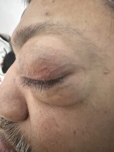
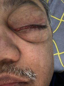
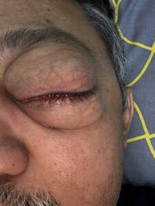
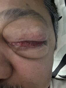
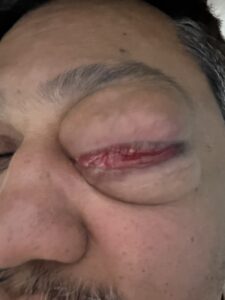
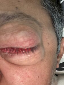
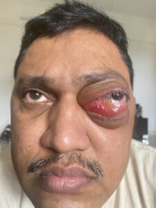
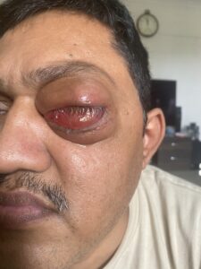
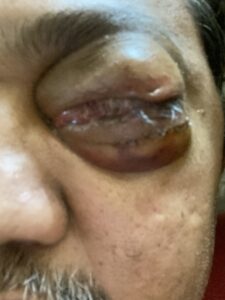
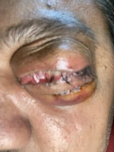
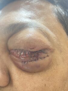
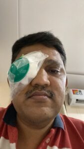
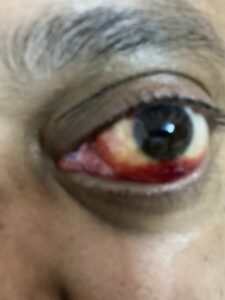
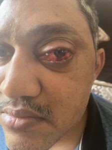
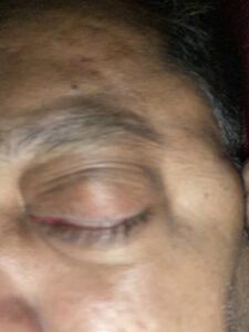
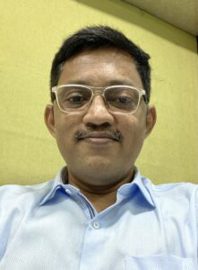
Photograph documenting the visible swelling/proptosis starting from November, 2022 to August 2026


PET/CT report image shared by me.
MRI report image shared by me.
सतत विचारले जाणारे प्रश्न
What is an atypical lipomatous tumour (ALT) of the orbit?
ALT is a rare fatty tumour. In difficult anatomical locations, it is closely related to well-differentiated liposarcoma (WDLPS). Diagnosis requires correlation of imaging, pathology and molecular testing.
Can an atypical lipomatous tumour occur behind the eye?
Yes. Rare lipomatous tumours can occur within the orbit, including intraconal and extraconal spaces. My lesion involved both compartments.
Is orbital ALT the same as well-differentiated liposarcoma?
ALT and WDLPS are essentially the same biological entity, with terminology influenced by anatomical site and resectability. An expert pathologist should make the final interpretation.
Is orbital liposarcoma a cancer?
Liposarcoma is a malignant soft-tissue tumour. Well-differentiated tumours generally behave less aggressively than high-grade sarcomas, but they can cause serious local problems and recur.
Can an orbital liposarcoma spread to the brain or sinuses?
It can extend locally if it progresses, but local extension is different from distant metastasis. My reported imaging did not show brain invasion.
Can orbital ALT spread to other parts of the body?
ALT/WDLPS generally has a low metastatic risk compared with dedifferentiated or high-grade liposarcoma, but recurrence and dedifferentiation remain important concerns.
What do MDM2 and CDK4 amplification mean?
MDM2 and CDK4 copy-number gains/amplification are characteristic molecular findings in many ALT/WDLPS tumours. My report detected gains but also recommended confirmation with an alternate method.
Why is MDM2/CDK4 FISH testing important?
FISH can directly assess gene amplification and may help resolve difficult lipomatous tumour diagnoses. The treating pathologist should decide the appropriate confirmatory test.
Can orbital ALT be treated without removing the eye?
In selected cases, eye-sparing treatment may be possible, but feasibility depends on tumour location, margins, optic-nerve involvement and extraocular muscle involvement. This requires expert orbital tumour assessment.
What is globe-sparing surgery?
It means trying to remove or control the tumour while preserving the eyeball. Whether adequate tumour control can be achieved this way is highly individual.
When is orbital exenteration recommended?
It may be recommended when a tumour extensively involves the orbit and cannot be adequately controlled with a less extensive operation. The decision should be based on imaging, pathology and surgical anatomy.
Can radiotherapy treat orbital ALT/WDLPS?
Radiotherapy can be useful for selected soft-tissue sarcomas, but its role in a particular orbital ALT/WDLPS must be assessed by a specialist radiation-oncology and sarcoma team.
Are medicines available for CDK4/MDM2-amplified liposarcoma?
CDK4/6 inhibitors and MDM2-directed therapies are being studied, especially in advanced or dedifferentiated liposarcoma. They are not established replacements for surgery in localized orbital ALT/WDLPS.
Can homeopathy or Ayurveda cure an orbital lipomatous tumour?
There is no established clinical evidence that homeopathy or Ayurveda can eradicate ALT/WDLPS. I report my experience for transparency, not as proof of efficacy.
Can an orbital tumour remain stable for months or years without surgery?
Some published cases have shown prolonged stability, but this cannot predict what will happen in another person. Continued specialist examination and imaging are essential if observation is chosen.
Search Questions and Keywords for Patients Looking for Similar Cases
- orbital atypical lipomatous tumour / ALT of orbit
- well-differentiated liposarcoma behind the eye
- orbital liposarcoma treatment
- fatty tumour behind eye / fat lump in eye socket
- left orbital mass with proptosis
- tumour involving medial rectus muscle
- orbital tumour compressing optic nerve
- MDM2 amplified orbital tumour
- CDK4 amplified liposarcoma
- MDM2 FISH for orbital tumour
- can orbital liposarcoma be removed without removing the eye
- globe-sparing surgery for orbital tumour
- alternative to orbital exenteration
- orbital exenteration for liposarcoma
- stable orbital liposarcoma
- orbital ALT surveillance
- targeted therapy for MDM2/CDK4 liposarcoma
- orbital liposarcoma clinical trials
- rare tumour behind eye
- rare eye socket tumour